Purpose of this website
This webpage provides a summary and installation instructions of software that may be used for filling white matter lesions on T1-w images, like multiple sclerosis (MS) lesions, in brain MRI.
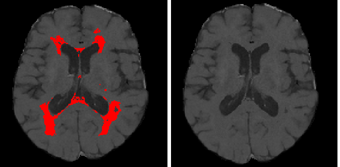
Filling the lesions is a crucial step in quantitative volumetric studies and analyses of brain atrophy. For instance, MS white matter lesions can affect brain tissue volume measurements of voxel-wise segmentation methods if these lesions are included in the segmentation process. It has been demonstrated that automatic tissue segmentation methods are more accurate when white matter lesions are filled with "synthetic white matter ". Over the last years, several lesion filling approaches have been presented, although none of them can be considered as a standard de facto algorithm. In this page we provide links to different algorithms and also a novel one to perform this task.
If you feel interesting our webpage and use some of the resources given, please cite the following paper in your research:
- S. Valverde, A. Oliver, X. Lladó. A white matter lesion-filling approach to improve brain tissue volume measurements. NeuroImage: Clinical, (6), pp 86-92, 2014.