Project reference: DPI2015-68442-R
Budget: 114.400 €
Duration: 3 years
Principal researcher: Dr. Robert Martí Marly
Breast cancer screening based on mammography (MG) has had a major impact on reducing mortality at affordable cost. However, overdiagnosis, overtreatment, and the lack of sensitivity of mammography, especially in women with dense breasts, need to be addressed to improve screening. More effective screening cannot be achieved with the generalised use of MG for all the population; personalisation is required where the density of a woman’s breasts or increased cancer risk is assessed prior to determining whether or not additional imaging (i.e. magnetic resonance imaging (MRI), 3D ultrasound (ABUS), or tomosynthesis (DBT)) is necessary. Hence, the question is no longer if the screening regimens will be personalised, but mainly how this can be efficiently implemented.
Although these complementary modalities are able to increase the sensibility compared to MG alone in specific patient groups, they still present several drawbacks that limit their effectiveness in personalised screening scenarios. Those are mainly related to technological issues linked to their novelty and to diagnostic accuracy, increased costs and reading times.
Researchers in the SMARTER project believe that image analysis techniques can be used for the development of Intelligent Diagnostic Enhancement tools based on automated detection and density estimation in ABUS, DBT and MRI. These tools will impact in improving reading times and diagnostic accuracy of these modalities in order to be used in personalised breast screening scenarios. Hence we aim to develop and evaluate novel imaging tools that can be integrated early into the screening workflow to steer image acquisition and guide the selection of appropriate personalised screening protocols; and to process imaging data in an intelligent way to minimise interpretation time. Tools will be based on breast density estimation algorithms and automated breast cancer detection algorithms applied to DBT, ABUS and MRI.
Challenges:
Breast Density
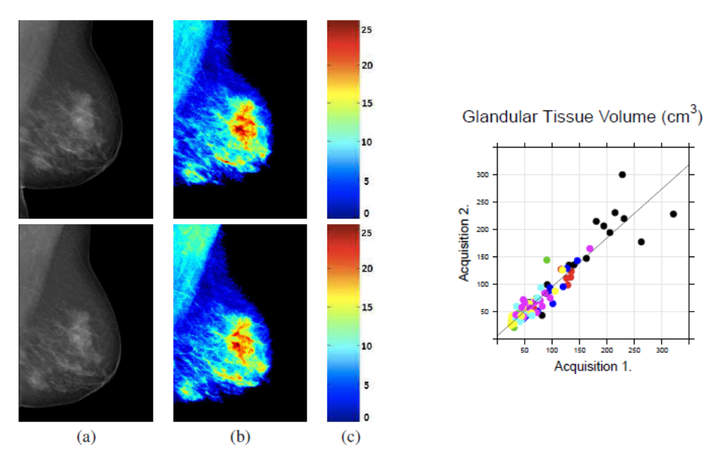
To develop methods for quantitatively assessing the breast density in DBT and MG, based on modelling the imaging physics and using image analysis techniques for breast tissue characterisation. Evaluation comparing to commercial software (Volpara) and other modalities (MRI) it is also investigated. Example of density estimation in MG and a methodology for density estimation in DBT.

Cancer detection
To develop methods for cancer detection and segmentation for MRI, MG, DBT and ABUS, in particular:
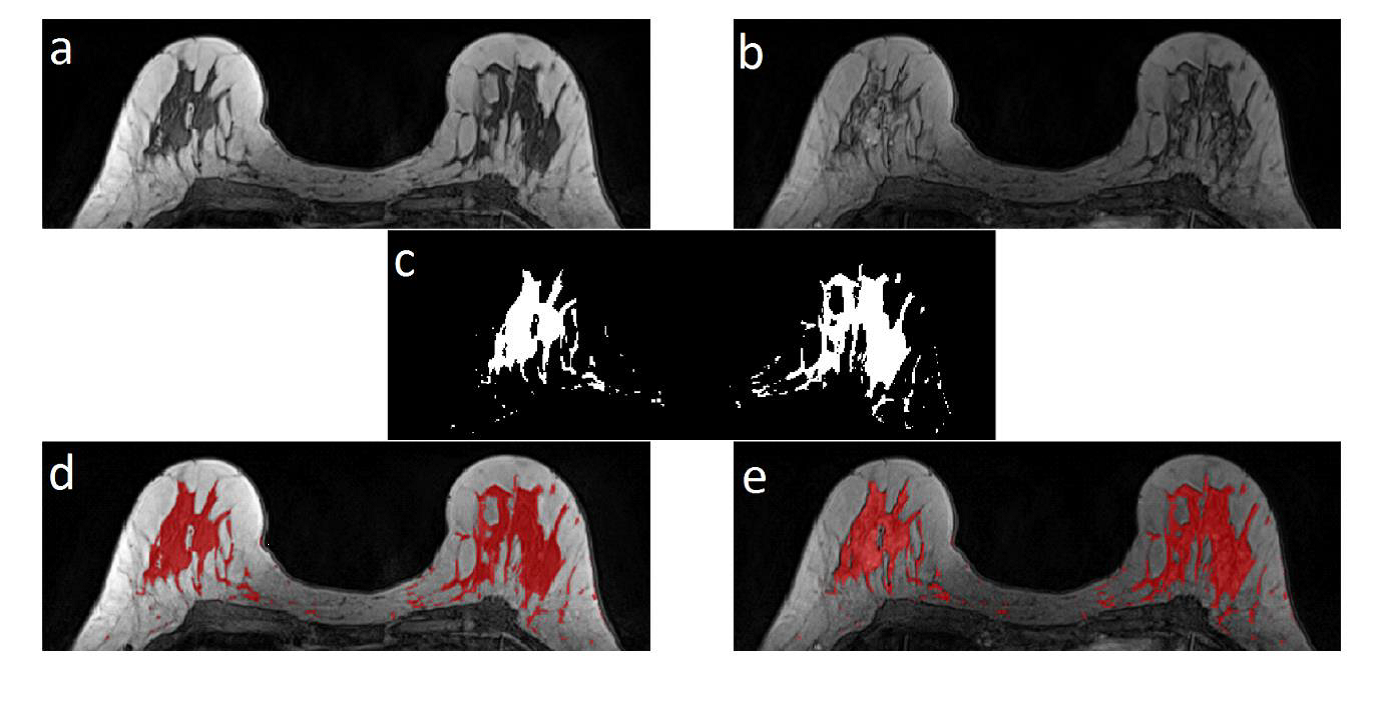
- To develop further evaluate methods for cancer detection in conventional MRI and to propose methods for quantitative analysis of breast parenchymal enhancement (BPE) for risk estimation and assessing treatment response. Example of BPE estimation in MRI.
- To develop methods for cancer detection and segmentation and study of the impact on the DBT acquisition parameters and systems in the accuracy of the system.
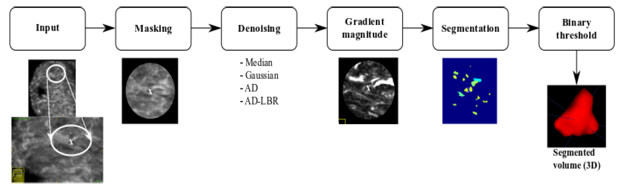
- To develop and evaluate cancer detection and segmentation and lesion follow-up in temporal studies to assess lesion changes over time in ABUS. The figure shows the lesion segmentation framework in ABUS